



Provide high-precision non-linear 3D registration.

Achieve high-speed calculation!
Due to the complexity and high cost of DIR calculations, the problem was the increased computation time. To address this, parallel computing with CPUs, GPUs, and CPU/GPU hybrid computing was implemented to achieve faster speeds. Additionally, the computation algorithm was optimized for further acceleration.

Innovative error estimation method
The error evaluation of DIR is most easily done through visual evaluation using difference images. While quantitative evaluations such as ICE (inverse consistency error) have been proposed, they assume that the error of the inverse transformation of image registration is infinitely small. However, this product developed its own error evaluation method to represent errors on the image, achieving both quantitative and visual evaluations.

Semi-automatic contouring
By reading in the ROI shape data of the reference breathing phase and inputting it into this product, automatic calculation of ROI shapes for other breathing phases can be achieved. Furthermore, with its excellent user interface, natural modification of ROI shapes can be performed.
How DIR works
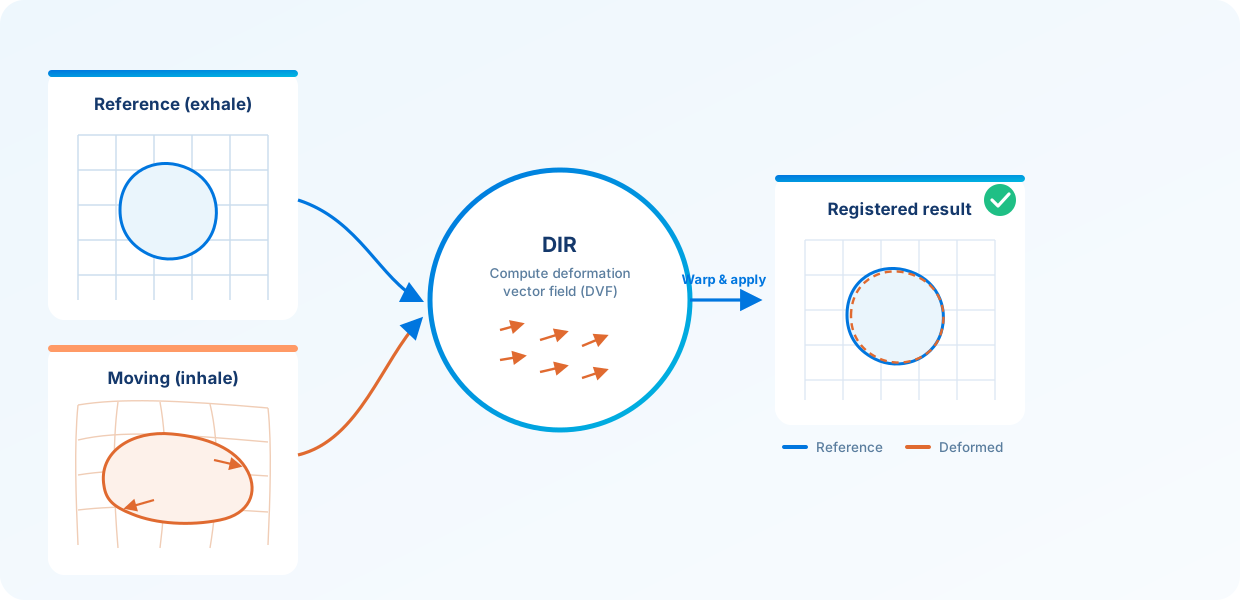
DIR (deformable image registration) accurately overlays two 3D images that have been deformed by breathing or changes in body position, taking the deformation into account. One image is used as the reference and the other as the moving image; the moving image is locally deformed to compute, at the pixel level, the deformation field (deformation vector field: DVF) that best matches the reference. Organ deformations that rigid registration—translation and rotation only—cannot resolve are corrected non-linearly by the DVF, achieving precise alignment.
An easy-to-read error map
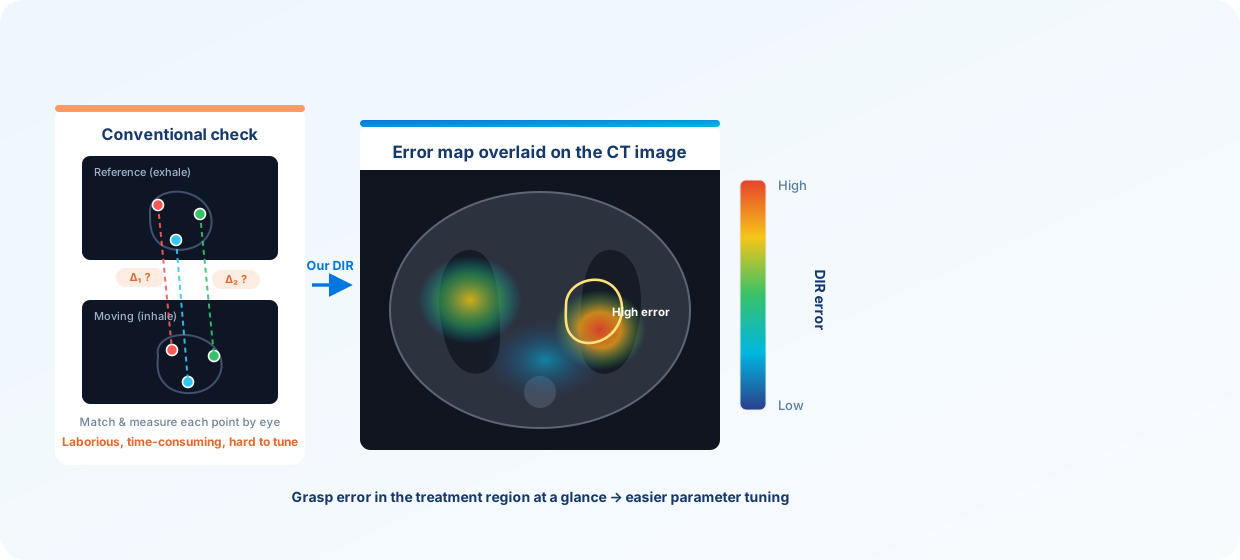
Evaluating DIR error is laborious and time-consuming, which makes tuning the optimal DIR parameters difficult. Our DIR overlays the error map directly onto the CT image so the error can be grasped at a glance. This lets you instantly check the DIR error in regions relevant to treatment and makes parameter tuning far easier.
An easy-to-use interface
We also provide an intuitive graphical user interface (GUI). Reviewing and editing ROI shapes, adjusting DIR parameters, and displaying the various results can all be done smoothly on screen. The design aims to be approachable even for users who are not accustomed to specialized operations.
Computing the time-integrated dose distribution
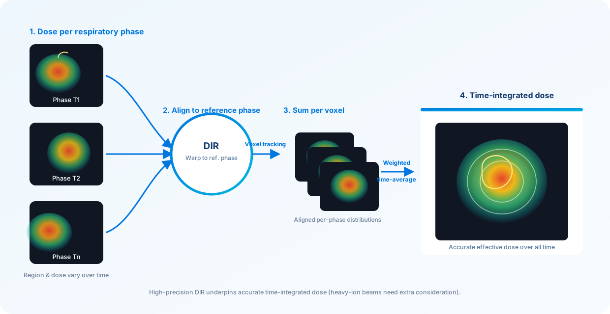
When the dose distribution is calculated for each respiratory phase using 4D CT images, some regions are irradiated and others are not depending on the time, and even the same region may receive a different dose at different times. The distribution at each time point alone does not allow a comprehensive dose evaluation. Therefore, the deformation of the CT image at each respiratory phase is computed by DIR and warped to the reference phase, so the same voxel can be tracked over time and its dose summed across all time points. A weighted average along the time axis then yields the time-integrated dose distribution (heavy-ion beams require additional considerations). Accurately computing such a dose distribution depends on high-precision DIR.